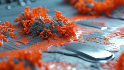
| Fibrillar Type I Collagen |
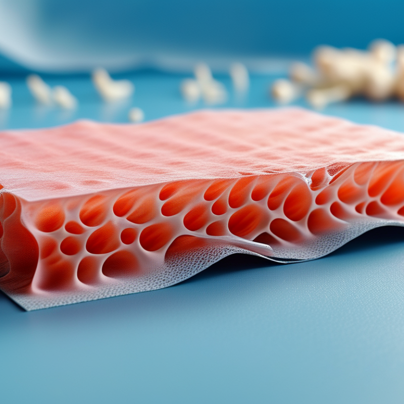
Purified fibrillar collagen is neutralized and gelled, then formed as a sponge, membrane, gel, or injectable matrix. It may be stabilized by physical or chemical crosslinking. |
Open porous sponges commonly use approximately 50–300 µm pores; pore size depends strongly on freezing, drying, solids concentration, and crosslinking conditions. |
Generally soft and hydrated. Useful for cell-contact environments, but native or lightly crosslinked forms have limited load-bearing capacity and can contract during culture. |
Fast to moderate enzymatic degradation. Crosslinking usually increases resistance to collagenase and reduces shrinkage, but excessive crosslinking can reduce remodeling. |
High cell-adhesion potential, natural integrin-binding sites, good support for fibroblasts, osteoblast-lineage cells, endothelial cells, and many stromal cells. |
Batch-to-batch variation, weak wet-state mechanics, possible contraction, and sensitivity to sterilization and storage conditions. |
Soft-tissue repair, wound coverage, cell culture matrices, injectable systems, and non-load-bearing regenerative products. |
| Atelocollagen |
Collagen with the non-helical terminal telopeptides removed to reduce some antigenic determinants. It can be processed into gels, sponges, films, or injectable formulations. |
Usually designed as a hydrated gel or porous matrix; pore dimensions are formulation-dependent and commonly fall within the tens-to-hundreds of micrometre range. |
Low intrinsic stiffness in hydrated form. Mechanical strength normally requires concentration adjustment, blending, or crosslinking. |
Typically more susceptible to enzymatic remodeling than heavily crosslinked collagen. Stability can be increased through covalent or enzymatic crosslinking. |
Reduced telopeptide-related immunogenicity compared with non-atelocollagen preparations and good biological recognition by adherent cells. |
Limited structural strength, potential rapid resorption, and sensitivity to concentration, pH, ionic strength, and temperature. |
Injectable matrices, dermal and soft-tissue regeneration, localized delivery, and cell-support formulations. |
| Collagen–Glycosaminoglycan Composite |
Collagen is combined with glycosaminoglycans such as chondroitin sulfate or hyaluronic-acid-derived components, commonly followed by freeze-drying and crosslinking. |
Interconnected pores are often engineered in the approximate range of 50–150 µm, although larger pores can be produced by changing freezing and drying parameters. |
Higher structural organization than many pure-collagen sponges, but wet-state strength remains moderate unless reinforced or strongly crosslinked. |
Degradation is adjustable through glycosaminoglycan content, crosslink density, and porosity. Greater crosslink density generally slows mass loss. |
Can reproduce aspects of extracellular-matrix composition, retain water, and support cell migration and matrix deposition. |
Hydration can reduce stiffness; glycosaminoglycan loss may alter pore structure and biological signaling during degradation. |
Skin, nerve, cartilage, tendon-interface, and other soft-tissue regeneration strategies where matrix composition is important. |
| Collagen–Hydroxyapatite Composite |
Mineral particles or nanoscale hydroxyapatite are incorporated into a collagen matrix, followed by molding, freeze-drying, mineralization, or additive manufacturing. |
Typical engineered macropores are often approximately 50–300 µm; nanoscale mineral domains are used to increase biomimicry and interfacial area. |
Higher compressive stiffness and improved osteoconductive character than collagen alone, but strength remains dependent on mineral fraction, orientation, porosity, and hydration. |
Collagen degrades enzymatically while the mineral phase dissolves more slowly and may remain as a calcium-phosphate reservoir. Degradation is formulation-specific. |
Combines collagen cell-interaction motifs with a mineral phase resembling bone extracellular matrix; supports osteogenic differentiation and mineral deposition under suitable conditions. |
Excess mineral can reduce flexibility and cell-accessible collagen; weak interfaces or nonuniform mineral distribution may cause brittle failure. |
Bone void filling, craniofacial repair, periodontal regeneration, and other mineralized-tissue applications. |
| Electrospun Collagen |
Collagen is electrospun into aligned or randomly oriented nanofibers. Stabilization or blending is commonly required because pure collagen fibers can be weak and water-sensitive. |
Fiber diameters are commonly in the 100 nm–2 µm range. Inter-fiber pores are often relatively small and may restrict deep cell infiltration without additional design features. |
Good fiber-level tensile behavior and anisotropy when aligned, but wet-state strength and dimensional stability depend strongly on crosslinking and fiber packing. |
Unstabilized collagen can degrade rapidly and lose fibrous architecture in aqueous environments; crosslinking improves persistence. |
High surface area, fibrillar topography, and controllable alignment can guide cell orientation and extracellular-matrix organization. |
Low thickness, limited cell penetration, solvent or processing concerns, and potential loss of native bioactivity after harsh stabilization. |
Tendon, ligament, nerve guidance, vascular-interface, skin, and barrier-membrane designs. |
| Collagen Hydrogel |
Neutralized collagen solution is gelled under physiological or near-physiological conditions. It may be delivered by injection or combined with cells, growth factors, or reinforcing fibers. |
Hydrogel mesh size is generally smaller than the macropores of freeze-dried sponges; effective transport depends on collagen concentration, fibril diameter, and gel density. |
Very soft and highly deformable. Suitable for non-load-bearing environments, but often requires reinforcement for handling and long-term shape retention. |
Usually rapid enzymatic remodeling unless concentration, fibril density, or crosslinking is increased. |
Excellent conformability, cell encapsulation potential, high water content, and minimally invasive delivery options. |
Low mechanical strength, contraction, syneresis, and possible difficulty maintaining a defined geometry after implantation. |
Injectable cell delivery, soft-tissue regeneration, localized factor delivery, and defect-filling applications. |
| Methacrylated Collagen |
Collagen is chemically functionalized with methacrylate groups and photocrosslinked or otherwise polymerized to create a tunable hydrogel or printed construct. |
Microscale network dimensions are controlled by collagen concentration and crosslink density. Printed architectures can include designed macropores from approximately 100 µm to 1 mm or more. |
Higher geometric stability and tunable modulus than uncrosslinked collagen hydrogels; stiffness increases with polymer concentration and light-induced crosslink density. |
More stable than uncrosslinked collagen, but hydrolysis and collagenase-mediated remodeling still occur. Degradation can be adjusted through substitution and crosslinking. |
Combines collagen-derived cell-interaction motifs with phototunable shape fidelity and spatial control over architecture. |
Photoinitiator cytocompatibility, light penetration, residual reagent removal, and possible reduction in cell-remodelable sites must be evaluated. |
Three-dimensional bioprinting, patterned tissue models, organoid support, and customized non-load-bearing constructs. |
| Collagen–Fibrin Composite |
Collagen is combined with fibrin to form a hydrated composite, often using thrombin-mediated fibrin formation or sequential gelation. |
Fine fibrillar networks are formed within a hydrated matrix; macropore size is generally determined by molding, porogen use, or secondary reinforcement. |
Fibrin can improve early handling and cell-mediated remodeling, but the composite is normally soft unless reinforced or crosslinked. |
Fibrin is usually remodeled relatively quickly, while collagen provides longer-lived structure. The balance depends on fibrin content and stabilization. |
Supports cell migration, vascularization-related studies, and dynamic matrix remodeling; both components provide biologically recognizable environments. |
Rapid shrinkage or contraction, variable clot structure, limited load-bearing capability, and sensitivity to enzymatic activity. |
Vascularized tissue models, wound healing, soft-tissue repair, and cell-laden regenerative constructs. |
| Decellularized Collagen-Rich Matrix |
Native tissue is decellularized to retain collagen architecture and selected extracellular-matrix components. The material may be used as a sheet, particulate matrix, hydrogel, or bioink. |
Architecture is tissue-specific and may range from aligned micron-scale fibers to interconnected pores from tens to hundreds of micrometres. |
Can preserve tissue-specific anisotropy and handling properties, but mechanical performance varies substantially with tissue source, processing, and sterilization. |
Remodeling is biologically active and source-dependent. Harsh processing may improve decellularization but damage collagen structure or remove signaling molecules. |
Retains tissue-specific biochemical cues, structural motifs, and matrix-bound components that may support constructive remodeling. |
Source variability, incomplete decellularization, residual immunogenic material, sterilization effects, and more complex quality control. |
Organ-specific soft-tissue models, wound repair, vascular or nerve interfaces, and advanced tissue-engineering research. |
| Aligned Collagen Bundle or Fiber-Reinforced Scaffold |
Collagen fibers, bundles, tapes, or aligned porous layers are assembled to reproduce directional tissue structure. They may be combined with a collagen gel or particulate phase. |
Fiber alignment is the primary design variable; bundle spacing and inter-bundle pores are selected to balance cell infiltration with directional load transfer. |
Improved anisotropic tensile behavior compared with random sponges. Performance depends on fiber continuity, crimp, hydration, crosslinking, and interface quality. |
Gradual enzymatic remodeling is possible, but aggressive crosslinking may delay turnover and reduce biological integration. |
Provides contact guidance and directional mechanical cues for cells and matrix deposition. |
More difficult manufacturing, nonuniform stress distribution, possible delamination, and limited suitability for isotropic defects. |
Tendon, ligament, meniscus, nerve, muscle-interface, and other directionally organized tissues. |
| Comparison note: Reported pore sizes, stiffness, degradation times, and biological outcomes are representative ranges or qualitative tendencies from collagen-scaffold research. They are not universal specifications. Final comparison should use the same collagen source, concentration, crosslinking method, sterilization process, hydration state, test temperature, loading mode, and endpoint definition. |
| 2026 selection priorities: Match scaffold architecture to the target tissue, then verify wet-state mechanics, mass transport, cell infiltration, degradation–regeneration timing, sterilization compatibility, residual crosslinker or photoinitiator levels, manufacturing reproducibility, and applicable regulatory requirements. |