2026 Best 3D Bioprinting Technology for Global Buyers

As 2026 approaches, global buyers are examining 3D bioprinting technology beyond impressive demonstrations. They want dependable systems that produce consistent results, support trained operators, and fit real laboratory workflows. A printer may create detailed tissue structures, yet performance depends on bioinks, cell handling, software, sterilization, and maintenance. The machine is only one part.

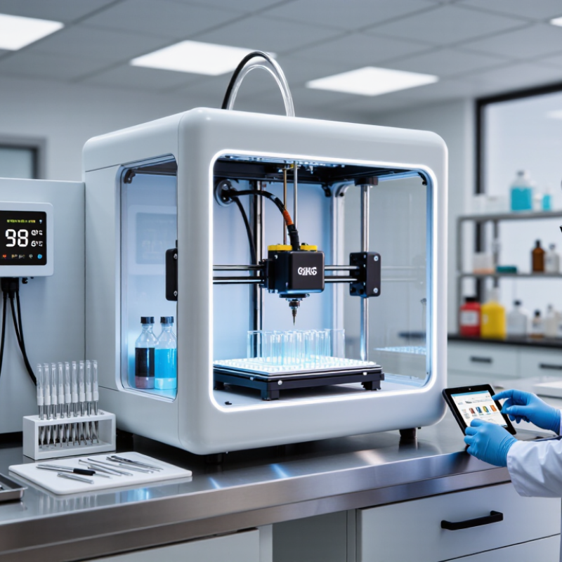
In research laboratories, experienced teams compare resolution, print speed, cell viability, material compatibility, and environmental control. They also review training requirements, service response times, consumable availability, and total ownership costs. These details become visible beside the printer, where calibration records, sterile tools, and temperature monitors shape daily outcomes. Strong suppliers provide technical documentation, transparent specifications, validation guidance, and evidence from credible studies or institutional users.

No platform wins everywhere. Some systems offer excellent precision but require expensive consumables or specialized staff. Others are easier to operate but provide fewer customization options. Buyers should question marketing language, request practical demonstrations, and examine performance under relevant conditions. Regulatory expectations also differ across regions, especially when systems support biomedical research or potential clinical development. Compliance cannot be assumed from a product brochure.

This guide evaluates leading solutions through a practical, evidence-informed lens. It considers current capabilities, application fit, supplier reliability, and long-term support for universities, biotechnology companies, hospitals, and industrial research teams. Results may vary between laboratories. That matters. A careful purchasing decision should balance innovation with reproducibility, safety, training, and honest recognition of technical limits.

2026 Best 3D Bioprinting Technology for Global Buyers

3D Bioprinting Technology: Definition, Principles, and Core Components

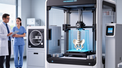
3D bioprinting is an additive manufacturing process that places living cells and biomaterials into organized, three-dimensional structures. Unlike conventional 3D printing, it must protect cell viability during printing. The technology often supports research into tissue models, drug testing, and regenerative medicine. It does not simply “print an organ.” That assumption needs careful correction.

The process begins with a digital model created from imaging data or computer-aided design. A printer then deposits bioink layer by layer, following programmed paths. Bioink may contain cells, hydrogels, growth-supporting materials, or combinations of these elements. Temperature, pressure, nozzle size, and printing speed directly affect cell survival and structural accuracy. Core components include the printhead, motion system, bioink reservoir, environmental chamber, software, and post-printing culture system. Each part influences the final result.

For global buyers in 2026, practical evaluation should include sterility control, calibration records, software compatibility, maintenance access, and operator training. Regulatory expectations also differ across markets, so documentation must be clear and traceable. A printed structure may look precise while cell distribution remains uneven. This is a real limitation. Buyers should request validation data, repeatability studies, and transparent performance boundaries. Faster printing is not always better. In some projects, a slower process protects delicate cells and produces more reliable research outcomes.

Major 3D Bioprinting Methods and Their Suitable Applications

For global buyers, the best 3D bioprinting technology depends on the tissue target, bioink, and required production scale.

Extrusion bioprinting is widely used because it handles cell-loaded hydrogels and viscous materials. It suits cartilage, bone scaffolds, skin models, and layered research constructs. A syringe deposits continuous strands, often resembling soft white threads on a culture plate. However, slow printing and nozzle stress may reduce cell survival.

Inkjet bioprinting places tiny droplets with high speed and reasonable positioning accuracy.

It works well for low-viscosity bioinks, drug testing platforms, and patterned cell studies. The method needs careful control of droplet size, temperature, and surface wetting. Small errors matter. A wet laboratory floor is not the issue; a slightly uneven cell pattern can change test results.

Light-based bioprinting cures photosensitive materials layer by layer, creating fine structures with smooth surfaces.

It can support vascular channels, organ models, and complex research scaffolds. Laser-assisted systems may position cells with excellent precision, but equipment costs, material limits, and operating expertise are higher. Buyers should request verified data on cell viability, sterility, resolution, maintenance, and regulatory suitability. Published performance is useful, yet real results can differ after changing cells, bioinks, or operators. No method fits every application. Careful pilot testing remains necessary.

Key Performance Criteria for Comparing Bioprinting Systems

For global buyers in 2026, comparing 3D bioprinting systems requires more than checking print speed. Resolution should match the intended tissue model, from fine vascular channels to larger scaffold structures. Ask for measured accuracy, not promotional estimates. Cell viability after printing is equally important. Request test data using relevant cell types, bioinks, and culture periods.

A reliable system should support repeatable layer thickness, stable temperature control, and consistent material flow. Throughput matters when experiments involve many samples, but faster printing can increase shear stress. Software usability also affects daily performance. Operators should be able to adjust pressure, speed, nozzle size, and path design without complicated workarounds. Sterile operation, cleaning procedures, calibration records, and technical training deserve equal attention. Small maintenance delays become expensive over time.

Tips: Build a comparison sheet with resolution, viability, printing volume, compatible bioinks, calibration frequency, and service response time. Test the system with your own materials before purchase. Speed alone misleads. Also inspect printed samples under magnification and measure actual dimensions. A perfect scorecard is unrealistic; one system may offer excellent precision but limited throughput. Experienced teams should document failed prints, too, because those results reveal operational weaknesses that polished demonstrations often hide. สล็อตโ

2026 Best 3D Bioprinting Technology for Global Buyers

Key Performance Criteria: Representative Minimum Feature-Size Ranges

Lower feature size generally indicates higher geometric resolution. The ranges shown are representative values reported across bioprinting research and technical reviews; actual performance depends on bioink properties, nozzle or optical configuration, layer height, crosslinking conditions, and tissue design. Global buyers should also compare cell viability, throughput, material compatibility, sterility, temperature control, and software workflow.

How Global Buyers Should Evaluate Suppliers and Compliance

For global buyers, the best 3D bioprinting technology is not defined by resolution alone. A reliable evaluation starts with the intended use, printed tissue type, and research setting. Ask suppliers for documented performance data, including cell viability, print repeatability, sterility controls, and material traceability. Request raw test conditions, not polished averages. This matters.

Supplier expertise should be visible in validation records, operator training, maintenance procedures, and change-control histories. Review whether the system supports controlled workflows, secure data handling, and environmental monitoring. Compliance evidence must match the destination market and the product’s risk classification. Check quality certificates, technical files, labeling, import documents, and post-sale reporting duties with qualified local advisers. A certificate alone proves little.

During demonstrations, observe cartridge loading, cleaning steps, software alerts, and time between print cycles. Ask how failed prints are investigated and recorded. Speak with existing users where possible, but verify their claims independently. Do not overlook service coverage, spare parts, cybersecurity updates, and staff turnover. I would question impressive data from small samples. That is uncomfortable. A lower purchase price can hide training gaps or delayed validation, creating higher ownership costs. Use a scored supplier matrix, retain evidence, and document every assumption before signing.

2026 Trends Shaping the International 3D Bioprinting Market

2026 Best 3D Bioprinting Technology for Global Buyers

The 2026 international 3D bioprinting market is moving toward smarter, more controlled production. Buyers increasingly seek systems that combine precise extrusion, gentle cell handling, and repeatable layer formation. Multi-material printing is gaining attention because tissues rarely contain one material or cell type. Improved bioinks are also supporting softer, more realistic structures for research and regenerative applications. However, faster printing does not always create better biological results. Cell survival, shape accuracy, and long-term stability still require careful testing.

Artificial intelligence is becoming a practical market trend, not just a promotional feature. Monitoring software can identify nozzle pressure changes, temperature shifts, and uneven layers during production. Cloud-based records may also help international teams compare batches across laboratories. Yet data quality remains a weakness. Poor calibration can produce confident but incorrect recommendations. Buyers should examine validation reports, maintenance access, training, and regional service capacity. Compliance requirements may differ between countries, so documentation must be clear and current.

Tips: Request sample data from real materials, not only demonstrations. Check sterility procedures and temperature control. Ask how failures are recorded and corrected. A small pilot project can reveal hidden costs, operator workload, and inconsistent results. Do not judge technology by resolution alone. Tissue performance matters more.

2026 Best 3D Bioprinting Technology for Global Buyers - 2026 Trends Shaping the International 3D Bioprinting Market

Technology Category Core Printing Principle Typical Feature Resolution Typical Layer Height Reported Cell-Viability Range Compatible Bioink or Material Classes Best-Fit Applications 2026 Buyer Outlook Main Limitation
Extrusion Bioprinting Pneumatic, piston-driven, or screw-driven deposition of continuous bioink filaments through a nozzle. Approximately 100–500 μm 100–500 μm Often 70–95% Hydrogels, alginate, gelatin-based systems, collagen, fibrin, decellularized extracellular matrix, thermoplastic polymers, and cell-laden composite inks. Tissue scaffolds, cartilage and bone models, skin constructs, organoid support structures, and research-scale regenerative medicine. High adoption potential because of broad material compatibility, relatively accessible operation, and scalable multi-nozzle configurations. Lower resolution and shear stress can affect delicate cells; printing speed and shape fidelity may decline as bioink viscosity increases.
Droplet or Inkjet Bioprinting Non-contact ejection of discrete droplets using thermal, piezoelectric, or acoustic actuation. Approximately 20–100 μm droplet scale 20–100 μm Often 80–95% Low-viscosity cell suspensions, growth factors, proteins, hydrogel precursors, and biomolecule solutions. Cell patterning, drug testing, tissue-interface models, microarrays, and localized biomolecule deposition. Strong for precision patterning and laboratory automation, especially where low material consumption is important. Usually requires low-viscosity materials and may face nozzle clogging, droplet instability, and limited structural strength.
Light-Based Projection Bioprinting Selective photopolymerization of a photosensitive bioink using projected visible or ultraviolet light. Approximately 25–100 μm 10–100 μm Often 80–95% Photocrosslinkable gelatin derivatives, polyethylene glycol-based hydrogels, methacrylated polymers, and other light-reactive formulations. Microstructured tissues, vascular-channel models, dental and cartilage research, organoid culture platforms, and complex soft-tissue geometries. High-growth precision segment due to fast layer exposure, smooth surfaces, and improved geometric resolution. Photoinitiator and light exposure must be carefully controlled; optical scattering, cytotoxicity, and limited material choices remain important concerns.
Laser-Assisted Bioprinting A pulsed laser transfers cell-containing droplets or bioink from a donor layer onto a receiving substrate. Approximately 10–100 μm 20–100 μm Frequently reported above 85% Cell suspensions, hydrogel droplets, proteins, and multiple biological materials deposited in precise patterns. High-resolution cell placement, skin and tissue-interface studies, microvascular research, and patterned co-culture models. Specialized premium segment for buyers prioritizing placement accuracy and heterogeneous tissue architectures. Higher system complexity, specialized consumables, more demanding alignment, and comparatively high capital and maintenance requirements.
Volumetric Bioprinting A complete three-dimensional structure is formed inside a photosensitive volume using multiple projected light paths. Approximately 100–300 μm Not layer-limited; approximately 100–300 μm effective detail Reported values commonly range from 70–95% Transparent or optically compatible photocurable hydrogels, cell-laden resins, and tissue-specific formulations under development. Rapid fabrication of soft-tissue geometries, embedded vascular structures, organoid holders, and complex research prototypes. Emerging 2026 technology with strong potential for reducing support structures and shortening print times for selected geometries. Limited resin transparency, light scattering, oxygen inhibition, material-development requirements, and less mature process validation.
Embedded or Support-Bath Bioprinting A soft bioink is deposited inside a temporary or permanent support bath that holds overhangs and freeform features. Approximately 100–500 μm 100–500 μm Often 75–95% Low-to-medium viscosity hydrogels, collagen-rich formulations, fibrin-based systems, and soft cell-laden bioinks. Tubular tissues, hollow organs, soft robotics for biomedical research, and structures with unsupported overhangs. Increasing research adoption where freeform geometry and low mechanical loading are more important than high throughput. Support-bath preparation and removal add process steps; contamination control and post-processing can be difficult to standardize.
Multi-Material and Multi-Cell Printing Two or more bioinks, cell types, or material chemistries are deposited within one construct using separate print channels or sequential processes. Approximately 50–500 μm, depending on the underlying method 50–500 μm Method-dependent; commonly 70–95% Hydrogels with different stiffnesses, cell-specific bioinks, conductive or mineral composites, and sacrificial materials. Heterogeneous tissue models, interfaces between hard and soft tissue, vascularized constructs, and disease-model platforms. One of the strongest 2026 trends because physiological tissues require spatially distinct cells, materials, and mechanical properties. Cross-contamination, nozzle calibration, bioink compatibility, synchronization, and higher validation workloads increase operational complexity.
AI-Assisted Process Control Computer vision, sensor feedback, and machine-learning models monitor filament width, layer placement, temperature, pressure, and defects. Depends on the printing platform; commonly 50–500 μm Platform-dependent Does not directly determine viability Compatible with extrusion, projection, droplet, laser-assisted, and hybrid systems. Repeatability improvement, automated quality inspection, parameter optimization, and reduction of failed research batches. Major international purchasing criterion as laboratories seek reproducible workflows, digital records, and lower operator dependence. Models require representative training data, validated acceptance criteria, cybersecurity controls, and human oversight.
Closed and Traceable Bioprinting Workflows Integrated sterile or controlled-environment printing, environmental monitoring, batch records, and standardized post-processing. System-dependent System-dependent Must be demonstrated by validated biological assays Sterile-compatible bioinks, single-use fluid paths, validated reagents, and controlled-temperature materials. Translational research, advanced therapy development, regulated laboratory environments, and reproducible preclinical testing. Growing priority for global buyers because documentation, contamination control, and repeatable manufacturing are increasingly important. Higher acquisition cost, consumable dependence, validation effort, and facility requirements.
Sustainable and Resource-Efficient Bioprinting Use of lower-waste deposition, reusable hardware where appropriate, reduced support material, and optimized energy and consumable management. Depends on the selected printing method Depends on the selected printing method Must be verified for each material and process Water-based hydrogels, bio-derived polymers, recyclable support media where technically suitable, and low-volume reagent workflows. Academic laboratories, high-throughput screening, cost-sensitive research centers, and institutions with environmental procurement requirements. Increasing procurement influence as buyers compare consumable waste, energy use, packaging, and total cost of ownership. Sustainability claims must be supported by life-cycle data; some low-waste materials may have lower printability or biological performance.
Data notes: Technical values are representative ranges reported in peer-reviewed bioprinting literature and technical reviews, not guaranteed specifications for every system. Cell viability varies with cell type, bioink chemistry, exposure conditions, nozzle size, pressure, temperature, post-print culture, and assay timing. Resolution refers to typical printed feature size or effective detail and should be confirmed through buyer-side validation.